

Imaging Options
X-Rays (Radiographs)
X-rays (or radiographs) are a staple of how we evaluate patients — they show bones, orthopaedic hardware (plates, screws, pins, replacements), and alignment of the bones and joints.
Depending on your condition, additional specialized x-ray views taken at specific angles can help us better understand your foot/ankle problem and treat you.
At HSS, patients typically get “weight-bearing” x-rays — that is, an x-ray taken while you are standing. This is often more helpful than a regular x-ray because it shows the alignment of your foot/ankle in a “functional” position.
Even if you have an MRI, CT scan, ultrasound or other imaging type, we will still obtain x-rays because they show these features (bones, hardware, alignment, functional position) so well.
X-rays do use radiation, but at very low doses. The average radiation dose from a standard set of x-rays in the foot/ankle is equivalent to the natural background radiation exposure you would get from just being on the planet for under 3 hours.

CT (Computed Tomography)
CT scans use radiation to provide 3-dimensional images of body parts, and they are especially good at looking at bones, especially fractures (broken bones), deformity/malalignment (crooked bones/joints), and healing of bones after a break or a surgery.
CT can be used used before total ankle replacement or other surgeries to create a customized, patient-specific surgical plan.
HSS is one of the first centers in the United States to use weight-bearing CT scans (WBCT), which shows the 3-D positions of the bones in a functional position.
Weight-bearing CT uses low radiation (up to 66% less than traditional CT).
You can watch a video of Dr. Henry’s discussion of technology and WBCT here, and read an article she wrote about the importance of WBCT in foot/ankle surgery here.

Watch Dr. Henry speak about WBCT in foot & ankle patients.
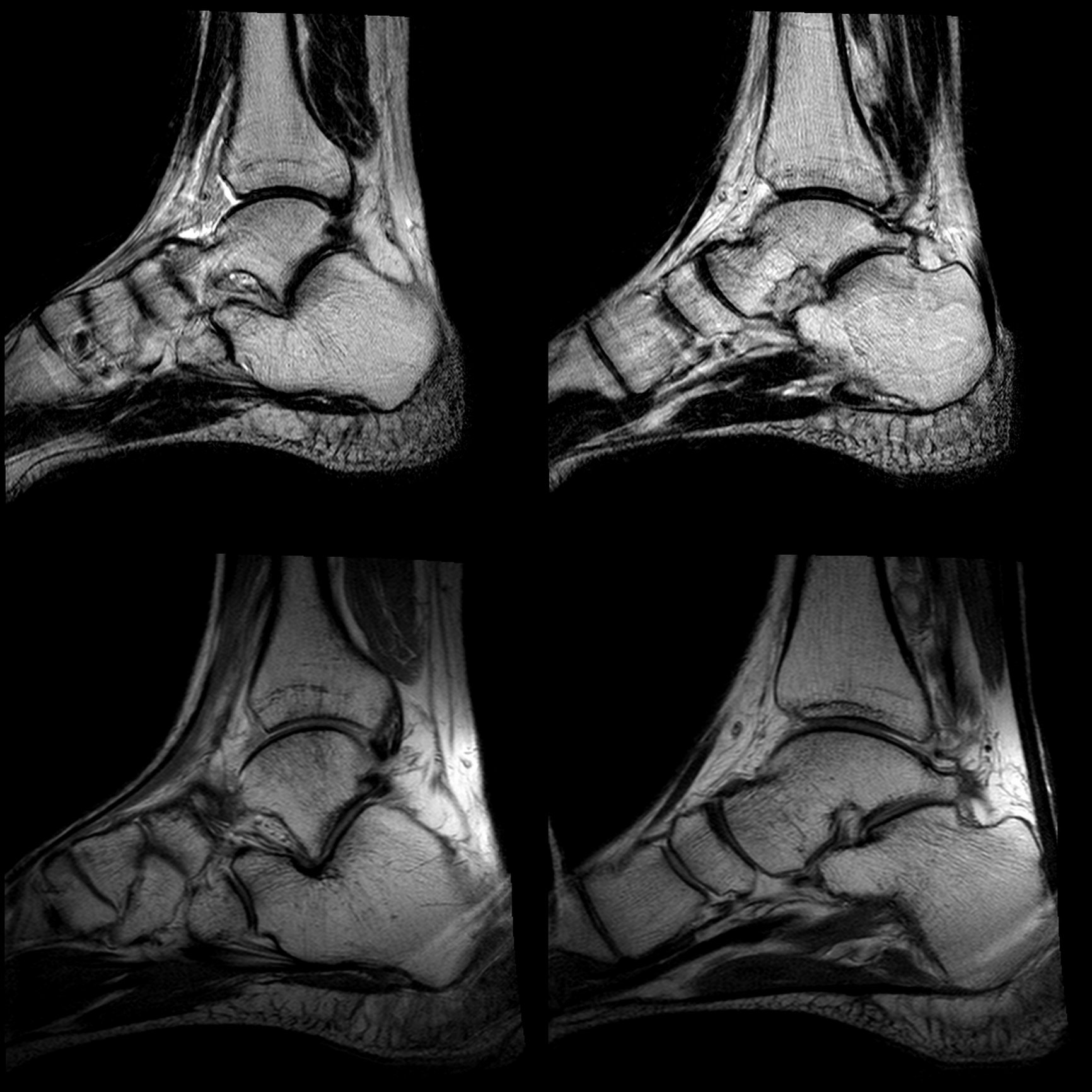
MRI (Magnetic Resonance Imaging)
MRI, or magnetic resonance imaging, helps us assess non-bony parts of the foot and ankle, including cartilage of the joints, tendons, and ligaments, as well as inflammation, or swelling, in tissues and bones — like in a “bone bruise” or other sprains/strains which often happen after trauma.
MRI does not use radiation to produce images.
However, MRIs take longer to perform (usually 30-60 minutes, depending on the study) and require you to lie still in the MRI during this time. The MRI scanner is non-weight bearing.
If you have metal or shrapnel in your body, or a pacemaker, discuss it with the radiology team prior to obtaining your MRI.
Ultrasound
Ultrasound uses a probe to that sends sound waves to the area of interest, which creates a picture of the area.
Ultrasound is safe (no radiation) and relatively easy to perform.
We can also use the ultrasound to look at tendons and ligament while the foot/ankle is moving, so we see what’s happening dynamically in real time.
However, the image quality is not as good as the others types, and it is helpful to have a radiologist/technique who is skilled/experienced with the technique.

Image-Guided Injections
If Dr. Henry recommends an injection of medication into a specific area, she may refer you to a radiologist to perform the injection under image guidance.
The radiologist will use x-ray or ultrasound to pinpoint the specific area, and inject the medication using the image to guide them in real-time. This confirms that the medicine is going to the actual site of the problem, and not a different area.
At HSS, there is a dedicated interventional radiology team who specializes in performing these types of injections, so they are very experienced and knowledgeable.
Other trained providers, including anesthesiologists, pain management physicians, and physiatrists, may also be able to provide image-guided injections.

The most important thing to know about imaging techniques is that they are all different, and they all provide different and valuable information. So, even if you have had an x-ray, a CT scan, or an MRI before, we may still recommend that we get one of the other studies to help better understand your foot/ankle problem. (Some patients with specific injuries may get all three.)
If you have any questions about why a certain image is being ordered, please ask. And if you are pregnant, trying to become pregnant, or unsure if you are pregnant, please let the office and radiology technicians know so that we can ensure your safety while still providing the best possible care.